Diagram Of The Muscles In The Forearm - Muscles Of The Anterior Forearm Flexion Pronation Teachmeanatomy
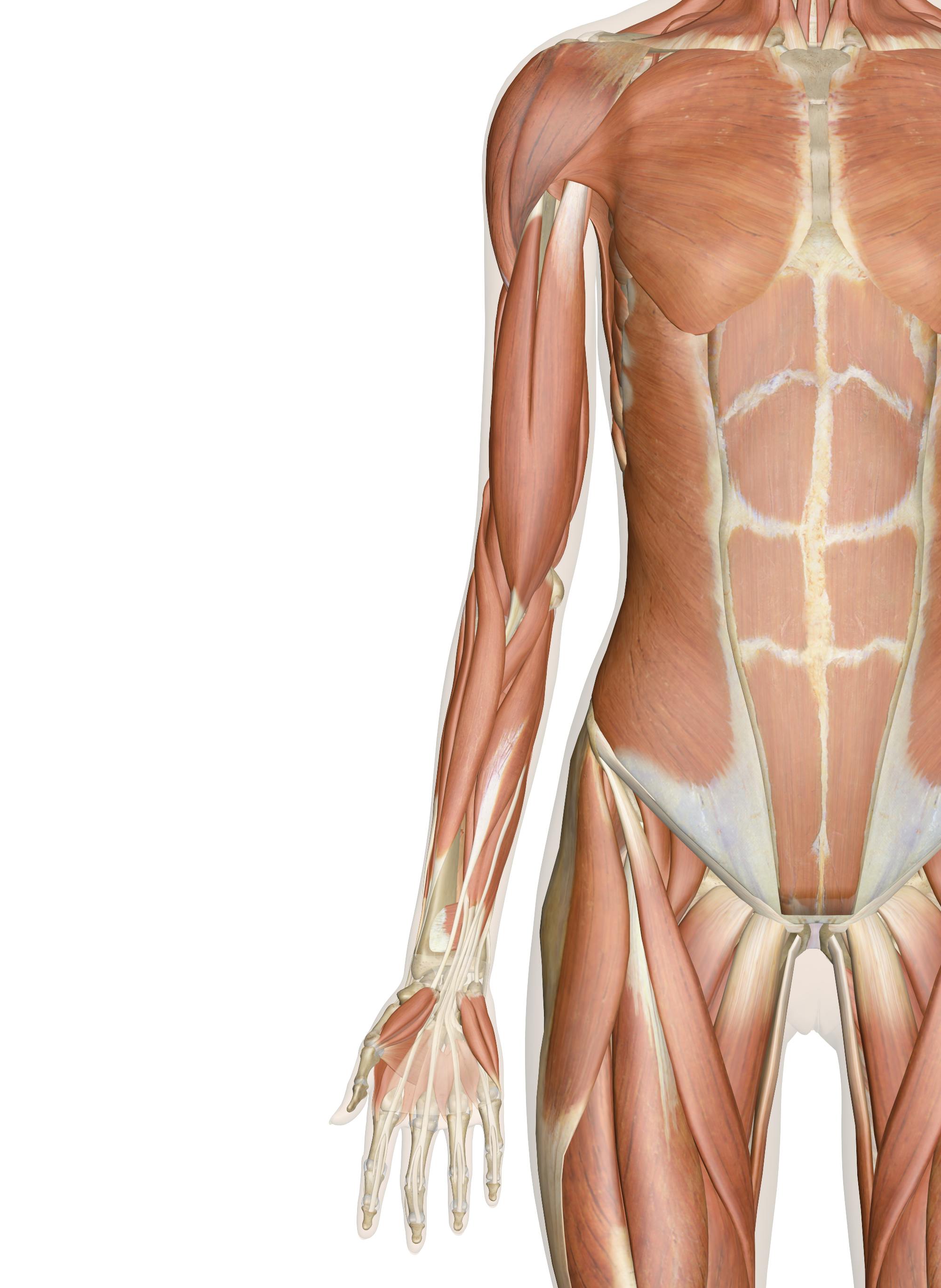
Diagram Of The Muscles In The Forearm - Muscles Of The Anterior Forearm Flexion Pronation Teachmeanatomy. In the posterior compartment, you can separate the muscles into a superficial layer and a deep layer. Superficial muscles of the posterior forearm: Pronator teres pronates the forearm, turning the hand posteriorly. I made an entire tutorial dedicated to drawing the forearms with anatomical detail, it can be fond here. The muscles of the upper arm are responsible for the flexion and extension of the forearm at the elbow joint.
ads/bitcoin1.txt
It arises from the grooved volar surface of the body of the radius, extending from immediately below. Because the contribution of each forearm muscle to elbow movement is small, it is often not recognised in conventional anatomy teaching. The forearm is the region of the upper limb between the elbow and the wrist. The forearm is a mass of some 20 different muscles. Here's an example of a petite woman.
Arm Muscles Anatomy Function Diagram Conditions Health Tips from post.healthline.com Diagram the movements of the humerus muscles that act on the forearm. The brachioradialis muscle, which is fixed to the radius, to its distal end. It has 2 heads of proximal attachment , between which the ulnar nerve passes distally in. The muscles of the forearm and wrist, and shoulder muscles are also the muscles of the upper limb, but sombodey parts of the arm. It occurs primarily in the articulation between the humerus and ulna and can achieve approximately 150° of movement. The anterior forearm muscles are divided into 3 muscular layers ; Learn vocabulary, terms and more with flashcards, games and other study tools. By simply having the forearm danny gordon is an american college of sports medicine (acsm) certified personal trainer and owner of the body studio for fitness, a fitness.
Here's an example of a petite woman.
ads/bitcoin2.txt
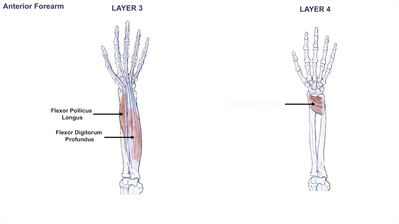
The muscles of this chapter are involved with motions of the forearm (radius and ulna) at the radioulnar joints, the hand at the wrist (radiocarpal) joint, and the fingers at the metacarpophalangeal (mcp) and/or the proximal. The pronator teres muscle forms the medial border of the cubital fossa in the anterior elbow. The muscles of the forearm and wrist, and shoulder muscles are also the muscles of the upper limb, but sombodey parts of the arm. Start studying muscles of the forearm. This layer contains only one muscle, the flexor digitorum. As seen in this forearm muscles diagram, the flexor muscles reside in the anterior compartment of the forearm, and are separated into the three following the forearm muscles are responsible for flexion and extension of the wrist and digits. Forearm flexion forearm flexion is rotation in the anatomic plane such that the radius and ulna move anteriorly. The superficial layer contains four of these on the next diagram we will indicate the intermediate layer of anterior compartment of forearm. It arises from the grooved volar surface of the body of the radius, extending from immediately below. Superficial muscles of the posterior forearm: Try labeling diagrams and worksheets as additional learning aids. A deep layer , intermediate layer and superficial layer. The flexor pollicis longus is situated on the radial side of the forearm, lying in the same plane as the preceding.
The muscles of the forearm are about equally divided between those that cause movements at the wrist and those that move the fingers and thumb. In the anterior compartment, they are split into three categories: The 3 muscle groups of the forearm each have their own unique form. This layer contains only one muscle, the flexor digitorum. Diagram the movements of the humerus muscles that act on the forearm.
Muscles Of The Arm And Hand Anatomy Pictures And Information from innerbody.imgix.net Muscles that participate in the same action, such as flexing the forearm, are actually partitioned off within the body into compartments by a tendinous sheathing called the intermuscular septum. Try labeling diagrams and worksheets as additional learning aids. By moving the mouse cursor over a particular area of the arm or forearm, this area is highlighted and the labels are displayed: Human muscle system, the muscles of the human body that work the skeletal system, that are under voluntary control, and that are concerned with the following sections provide a basic framework for the understanding of gross human muscular anatomy, with descriptions of the large muscle groups. There are many muscles in the forearm. Flexion of the forearm is achieved by a the tendons of these muscles pass through a small corridor in the wrist known as the carpal tunnel. Start studying muscles of the forearm. The antibrachial or forearm muscles may be divided into a volar and a dorsal group.
The antibrachial or forearm muscles may be divided into a volar and a dorsal group.
ads/bitcoin2.txt
The muscles of the forearm and wrist, and shoulder muscles are also the muscles of the upper limb, but sombodey parts of the arm. The anterior forearm muscles are divided into 3 muscular layers ; The brachioradialis muscle, which is fixed to the radius, to its distal end. I've just switched over to a diagram to show you this muscle. It is a functionally important muscle that contains two heads. Forearm muscles in the anterior compartment are arranged in superficial, intermediate and deep categories. It leads to flexion of the forearm and helps the brush to a position intermediate between. In the anterior compartment, they are split into three categories: This is the most medial of the superficial flexor muscles in the forearm. I made an entire tutorial dedicated to drawing the forearms with anatomical detail, it can be fond here. The antibrachial or forearm muscles may be divided into a volar and a dorsal group. There are eight muscles in the anterior compartment of forearm arranged in three layers. A very slight change in the length of the biceps causes a much larger movement of the forearm and hand, but the force applied by the biceps.
Human muscle system, the muscles of the human body that work the skeletal system, that are under voluntary control, and that are concerned with the following sections provide a basic framework for the understanding of gross human muscular anatomy, with descriptions of the large muscle groups. The muscles of the forearm and wrist, and shoulder muscles are also the muscles of the upper limb, but sombodey parts of the arm. The muscles of the upper arm are responsible for the flexion and extension of the forearm at the elbow joint. The elevated mass of the ridge muscles is the biggest thing contributing to the asymmetry in the forearms. In the anterior compartment, they are split into three categories:
Anatomy Of The Forearm Muscles And Tendons Lesson 1 Youtube from i.ytimg.com It occurs primarily in the articulation between the humerus and ulna and can achieve approximately 150° of movement. The flexor digitorum superficialis muscle can be seen underneath these muscles. The forearm is a mass of some 20 different muscles. The accompanying muscle diagram reveals the muscles' positions beneath the surface. A deep layer , intermediate layer and superficial layer. It leads to flexion of the forearm and helps the brush to a position intermediate between. There are many muscles in the forearm. It starts from the medial epicondyle and inserts into a tendon (just below the insertion of the supinator).
The 3 muscle groups of the forearm each have their own unique form.
ads/bitcoin2.txt
It arises from the grooved volar surface of the body of the radius, extending from immediately below. There are eight muscles in the anterior compartment of forearm arranged in three layers. The 3 muscle groups of the forearm each have their own unique form. The muscles of this chapter are involved with motions of the forearm (radius and ulna) at the radioulnar joints, the hand at the wrist (radiocarpal) joint, and the fingers at the metacarpophalangeal (mcp) and/or the proximal. Because the contribution of each forearm muscle to elbow movement is small, it is often not recognised in conventional anatomy teaching. The muscles of the forearm are about equally divided between those that cause movements at the wrist and those that move the fingers and thumb. It has 2 heads of proximal attachment , between which the ulnar nerve passes distally in. Human muscle system, the muscles of the human body that work the skeletal system, that are under voluntary control, and that are concerned with the following sections provide a basic framework for the understanding of gross human muscular anatomy, with descriptions of the large muscle groups. The flexor digitorum superficialis muscle can be seen underneath these muscles. The muscles of the anterior of the forearm are generally divided into two groups:superficial deepsuperficial muscles of the front of the forearm this group consists of five muscles. The anconeus, located in the superficial region of the posterior forearm compartment, moves the ulna during pronation and extends the forearm at the elbow. The forearm is the region of the upper limb between the elbow and the wrist. There are many muscles in the forearm.
ads/bitcoin3.txt
ads/bitcoin4.txt
ads/bitcoin5.txt
0 Response to "Diagram Of The Muscles In The Forearm - Muscles Of The Anterior Forearm Flexion Pronation Teachmeanatomy"


0 Response to "Diagram Of The Muscles In The Forearm - Muscles Of The Anterior Forearm Flexion Pronation Teachmeanatomy"
Posting Komentar